I ask the question “Is wear normal?” at almost every lecture I do on occlusion. Usually, the response is a small number of mumbled replies.
A good follow-up question is “How many eighty-five-year-old patients have you seen with mamelons?” I hope you are thinking not many if any at all.
So, yes, tooth wear of some amount is normal. A combination of attrition, erosion, and abrasion causes all of us to lose enamel over a lifetime.
Is the Wear Advancing at a Pathological Rate?
The more important question is when is the wear age-appropriate and when is it advancing at a pathologic rate?
We don’t have the data to know how many millimeters of enamel loss is appropriate for every decade of life. In order to help with this answer in my office, I play a mental game.
With the picture of the patient’s current wear in mind and a knowledge of their age, I imagine if the wear continues at the same rate at what age their teeth will be in jeopardy or need restorative dentistry to be saved.
I then reveal this estimate to the patient.
3 Ways To Document Wear Over Time
I believe it is important that I help my patients understand the process and the options for protecting their teeth.
1. Repeat Measurements
To quantify the amount of wear that is happening, we take a measurement from the CEJ to the incisal edge of several teeth with wear.
We take the measurement on the mid-facial and record it on the patient’s perio chart.
At subsequent appointments, we can now repeat these measurements and have clear data that the process is continuing.
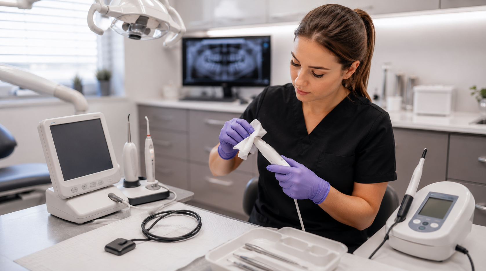
2. Repeat Photography
Another great way to document tooth wear is with photography.
With repeat photographs, we and the patient can see the change over time.
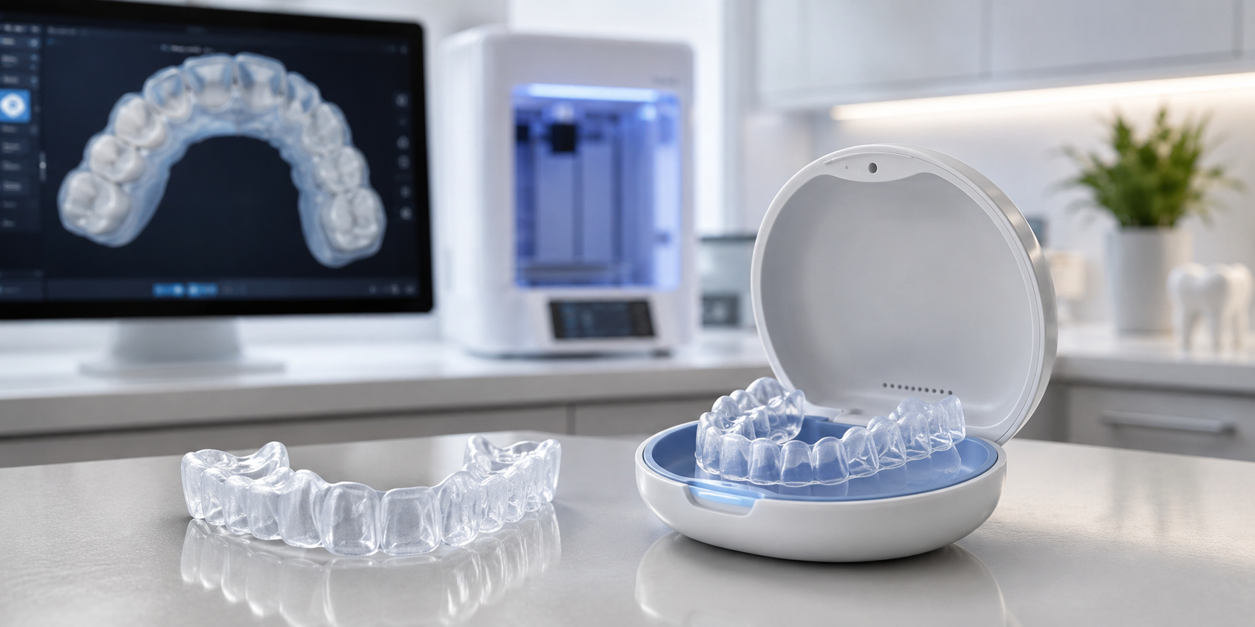
3. Repeat Digital Impressions
Today with digital impressions and software we can scan the arch, and then compare scans months or years later and get a precise measurement of the change.
What is Causing the Wear?
I believe that some wear is normal. I base this on the fact that I have very few if any patients who are in their seventies or eighties and still have mamelons on their incisors.
Wear is a concern when the amount of tooth structure being lost is outpacing the patient’s age.
I wrote about determining when wear leaves the physiologic category and becomes something we need to discuss with patients.
Both attrition and erosion can cause severe tooth wear, but they pose different long-term risks.
Once we have a sense of the cause of tooth wear, we can partner with the patient to treat the damage and manage the progression.
Guidelines for Discerning Attrition from Erosion
Attrition is the loss of tooth structure caused when the patient rubs two tooth surfaces together.
You will observe:
- Matching facets on upper and lower teeth
- Facets on tooth surfaces that occlude
- Enamel and dentin worn evenly
Erosion Caused by the Presence of Acid from Issues like GERD and Eating Disorders
You will observe:
- Facets that may or may not match on upper and lower teeth
- Facets on tooth surfaces that are not in occlusion
- Dentin cupped out and wearing faster than enamel
- Tooth structure wearing around restorations that remain unchanged
Note that attrition can be seen in addition to erosion, often giving us a false sense of how much the patient truly parafunctions, as the etched tooth structure wears away more easily.
Occlusal Therapy
Why Occlusal Appliance Therapy Is My First Step Prior to Ortho, Equilibration, or Restorative
Occlusal changes on an appliance are easy and reversible. An appliance can immediately reduce elevator muscle activity and give the patient relief.
The patient also experiences what changes to their tooth contacts could provide for them long term. We can test the changes that would be made by ortho, equilibration, and/or restorative.
Our goals are to stabilize the joint anatomy and reduce the activity of the elevator muscles because those muscles are what overload the joints and teeth.
We also want to slow down the rate of damage to the dentition and move that rate back to a more age-appropriate pace. We also may need to reorganize a patient’s occlusion to manage occlusal forces to ensure restorations that last.
Removing Posterior Contacts Does Not Work for Every Patient
Over my years of clinical practice, I have found that changing the occlusion does reduce functional risk for most patients.
However, we all have patients with perfect occlusion who present with TMD symptoms. We have some patients who continue to parafunction after we move them into immediate posterior disclusion.
Studies show that proprioception causes the elevator muscles to engage in only 80 to 85% of the population. This means that when the brain receives the signal that teeth are touching, the brain elevates the masseter muscles in 80 to 85% of people. Tooth contact is the trigger. Because this proprioception does not occur for 15 to 20% of the population, it is not the universal trigger for excessive loading.
Over my years in clinical practice, I have learned there is nothing I can do that is 100% dependable to stop a patient from para-functioning. Some of my patients continue to excessively load after posterior contacts are removed. Their functional risk does not diminish.
If we cannot reduce elevator force and redistribute force enough on an occlusal appliance to eliminate or at least relieve TMD symptoms, then occlusal therapy via ortho, equilibration, or restorative will not satisfactorily help the patient. We will need to turn to other forms of therapy.
Other modalities I use are BOTOX to deactivate muscles, massage therapy, and physical therapy. There are also systemic medications, cold lasers, and TENS therapy we can use to reduce the activity of the muscles or reduce inflammation in the muscles and joints. Sometimes one modality will alleviate symptoms for a while and when symptoms return, we can try it again or try another modality.
An Exercise to Identify the Patients Who May Not Benefit from Occlusal Therapy
You can do what I call a poor man’s EMG on yourself by placing your hands on your masseter muscles. Put your back teeth together, clench and release, clench and release, clench and release to see how much masseter activity you have. Then move your teeth into protrusive edge to edge and try to clench a little bit, making sure your back teeth do not touch. If you now have a posterior tooth touching in the edge-to-edge position, then put a pencil or pen between your front teeth to separate your back teeth.
With no back teeth touching and contact on the centrals, try to clench and release two or three times while feeling your masseters.
Most of you will find your masseters do not move or move a lot less when no back teeth are touching. Some of you, even with your back teeth separated, can still clench in protrusive and can still increase the muscle activity almost the same amount as when your back teeth touch.
I do this exercise with my patients, but when they move into protrusive, I put a bite stop over their front teeth or have them bite on a Lucia jig we have lined for their bite registration. If you do this test with your patients, you can use an EMG or feel the muscle activity with your hands.
If the patient can still generate almost the same force or the same force with their back teeth separated, you have identified one of the around 15% of people who might not benefit significantly from occlusal therapy.
You’ve also identified someone who might not do well on an anterior-only appliance because, if they can generate that same force on just two teeth, they are at risk for those teeth becoming sore and moving.
Interested in Learning More?
Join me at The Pankey Institute with The Essentials Series.
Starting with Essentials 1, this series offers a comprehensive and structured approach to dental education.
Participants will gain essential knowledge and skills, enabling them to build a solid understanding of fundamental concepts in dentistry.
From fundamental principles to essential clinical techniques, this series lays the groundwork for a successful dental practice and provides a strong platform for further specialization.