")
In esthetic dentistry, we spend a tremendous amount of time discussing tooth shape, color, and incisal edge position.
This is the third article in my photography series on smile design and facially driven dentistry. In the first article, we discussed why photography is essential in modern practice. In the second, we focused on how I use photography to determine incisal edge position. Now I want to discuss another critical layer of smile design that photography allows us to evaluate more objectively.
Gingival position and axial inclination are two of the most overlooked variables in esthetic dentistry, yet they dramatically influence whether dentistry feels harmonious or artificial.
And once again, photography becomes one of the most powerful tools we have to evaluate them objectively.
In my practice, I rely heavily on straight on retracted photos to analyze both gingival architecture and axial inclination before I ever propose a treatment plan. If these relationships are incorrect, even beautiful dentistry can still look wrong.
Photography Reveals What We Miss Clinically
When we are seated chairside, it is easy to focus only on individual teeth. Photography allows us to step back and evaluate the entire composition.
Once an image is placed on a large screen, patterns immediately become more obvious. Gingival asymmetries, incorrect zenith position, improper tooth angulation, discrepancies in axial inclination, uneven emergence profiles, and inconsistent connector progression become significantly easier to recognize photographically than clinically in real time.
Photography slows everything down and allows us to evaluate with intention rather than emotion.
Assessing Gingival Position
One of the first things I evaluate in a retracted smile photograph is the position of the gingival margins.
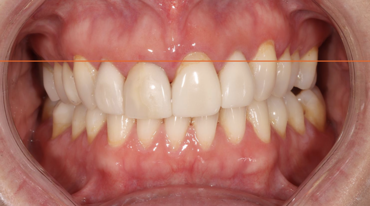
In an esthetically pleasing smile, the gingival architecture typically follows a predictable pattern. The centrals and canines generally share a similar gingival height, while the lateral incisors sit slightly more incisal. When that relationship is violated, the smile can begin to feel visually unbalanced even if the patient cannot immediately identify why.
This becomes especially important in restorative dentistry because we are often replacing or modifying teeth that already have altered gingival relationships.
Photography allows us to draw horizontal reference lines and evaluate symmetry objectively. Small discrepancies that may go unnoticed clinically become extremely obvious in a photograph.
In many cases, this becomes the determining factor in whether crown lengthening, orthodontic intrusion, extrusion, or restorative modification is necessary before finalizing treatment.
Gingival Zenith Position
Beyond overall gingival height, I also evaluate the position of the gingival zenith. The zenith is not centered on every tooth.
On maxillary centrals and canines, the zenith is typically positioned slightly distal to the vertical midline of the tooth. Laterals are often more centered.
When these positions are incorrect, the tooth can appear rotated, narrow, wide, or visually off balance even if the restoration itself is technically excellent.
Photography makes this significantly easier to evaluate because we can visualize the long axis of the tooth relative to the gingival architecture. This becomes especially important in veneer and anterior crown cases where subtle esthetic discrepancies are magnified.
Axial Inclination and Tooth Angulation
The second major parameter I evaluate photographically is axial inclination.
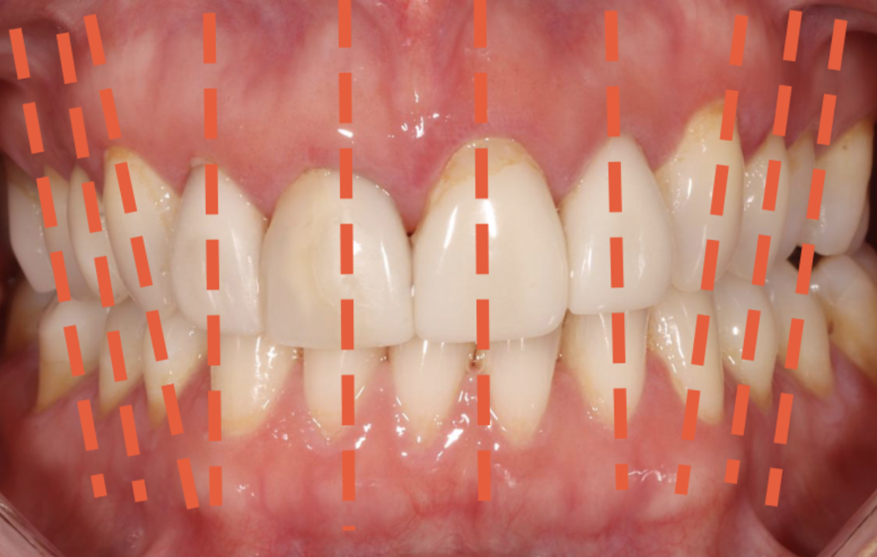
Axial inclination refers to the angulation of the long axis of the tooth relative to the facial midline and surrounding dentition. This has a tremendous influence on how natural a smile appears.
As we move posteriorly from the central incisors, there should typically be a progressive increase in mesial inclination. When that progression is absent, teeth can appear upright, flat, or artificial. When excessive, the smile can feel collapsed or crowded.
Photography allows us to evaluate these inclinations objectively by drawing vertical reference lines through the long axis of each tooth. Once these lines are placed, discrepancies become obvious immediately.
A single tooth with improper axial inclination can disrupt the harmony of the entire smile.
Why This Matters Restoratively
One of the biggest mistakes in esthetic dentistry is attempting to solve positional problems restoratively alone.
If the gingival position is incorrect, simply changing tooth shape rarely fixes the problem.
If axial inclination is incorrect, adding porcelain alone may actually exaggerate the issue.
Photography helps us determine whether the solution is restorative, orthodontic, periodontal, or most commonly interdisciplinary. I can then use these photos to communicate with specialists, allowing us to work together to execute the case.
Photography Creates Objectivity
The greatest value of photography in esthetic dentistry is objectivity. It removes the pressure and pace of chair-side diagnostics, allowing us to critically evaluate our work.
When we can objectively assess gingival position and axial inclination before treatment begins, we dramatically improve our ability to create dentistry that feels natural, balanced, and harmonious.
Because in the end, exceptional esthetic dentistry is rarely about one dramatic change.
It is usually the result of recognizing and controlling dozens of subtle relationships that most people never consciously notice, but immediately feel when they are correct.
Keep Reading: Incisal Edge Position