Occlusal Files
The Index Technique: New No-Prep Approach for Worn Dentition
By: Riccardo Ammannato, DDSThis Topic Originally Appeared on PankeyGram.Org. Dr. Ammannato granted permission for igniteDDS to share…

4 Main Reasons I Always Use Desensitizers Before Tooth Prep
By: Lee Ann Brady DMDTopic Originally Appeared on PankeyGram.Org. Dr. Brady granted permission for igniteDDS to share…
A Six-Unit Anterior Bridge: Provisionals and Placement
By: Lee Ann Brady DMDTopic originally appeared on Pankey.org: Dr. Brady allowed permission for igniteDDS to share…

Active Listening Tips For Patient-Centered Care
By: Dr. Bill Gregg DDSThis topic originally appeared on Pankey.org. Dr. Gregg granted permission for igniteDDS to…
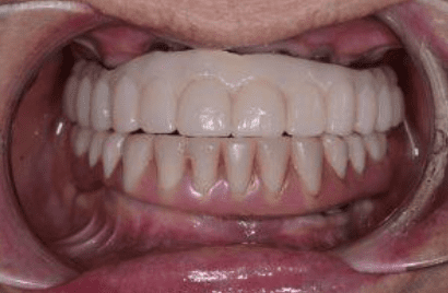
A Challenging Hybrid Overdenture Case
By: Dr. Lee Ann Brady, DMDThis topic originally appeared on Pankey.org. Dr. Brady granted permission for igniteDDS…
How to Master a Complete Dental Examination
By: Dr. Leonard A. Hess, DDS, Clinical Director, The Dawson AcademyThis article originally appeared on TheDawsonAcademy.com, Dr….

Shifting ‘The Locus of Control’ Towards a True Partnership
By: Brad WeissThis topic originally appeared on Pankey.org. Dr. Weiss granted permission for igniteDDS to share with…

Individualized Patient Details for Lifelong Relationships
By: DeAnne Blazek DDSThis topic originally appeared on Pankey.org. Dr. Blazek granted permission for igniteDDS to share…

What is Function and Aesthetics in Dentistry?
By: Dr. Neeraj KhannaTopic originally appeared on TheDawsonAcademy.com. Dr. Khanna granted permission for igniteDDS to share with…

Going Beyond Traditional Dentistry To Improve Dental Care
By: Bill Gregg DDSTopic originally appeared on Pankey.org. Dr. Gregg granted permission for igniteDDS to share with…