By: Jeffrey W. Horowitz, DMD, FAGD, D-ABDSM, D-ASBA
Have you ever run into facial asymmetries and discrepancies during your time as a dentist?
Here is a mini presentation on how I dealt with this.
Facial Asymmetries and Discrepancies: Case Study
Patient A:
- 13-year-old white female presents for an initial orthodontic evaluation
- Medical history is positive for occasional snoring, mouth breathing, and seasonal allergies.
- Dental/Perio Exam negative for pathology.
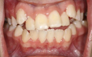
- Archform is narrow with a high palatal vault.
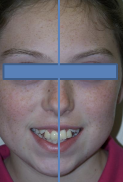
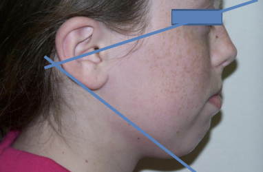
- Skeletal Class II with anterior and left-sided open bite and strained lip seal.
- Very high mandibular plane angle.
- Lower midline discrepancy.
- Ectopic eruption of Mx Canines.
- Occasional audible “Pop” in right TMJ.
- Soft tissue exam positive for slight tonsillar hypertrophy.
- Poor tongue posturing was noted.
Etiology of Facial and Dental Asymmetry
1. Foundational problems/injuries at the level of the TMJ, resulting in disk pathology, growth disruption or arthritic changes. Foundational injuries will typically create a more class II appearance on the side of the injury with the movement of the mandibular midline also skewed toward the altered foundation.
2. Injury or growth disruption of the facial bones.
3. Dental asymmetries can exist for many reasons including missing teeth, delayed eruption patterns, ectopic eruption retained deciduous teeth, tongue/oral habits, airway discrepancies, and foundational injuries
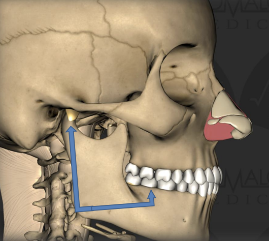
Patients with foundational discrepancies may be at higher risk for airway obstruction.
Taking the “Functional 4” Photos Can Reveal Problems
1. Full Face Lips Together
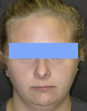
2. Full Face Smile
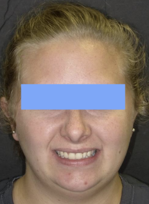
3. Profile Lips Together
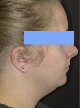
4. 1:2 Retracted MIP
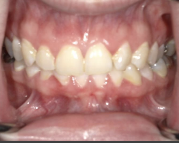
Back to Patient A
Despite the difficulty of the case, airway, and TMJ risks, straight-wire orthodontics were initiated to correct class II occlusion and close open bite…MISTAKE!
The treatment extended over 3 years and despite bite closure mechanics, could not close the bite. Occlusal prematurities prevailed on the posterior right.
A late-treatment CBCT (did not have technology at the onset of the case) showed arthritic changes to the right condylar head, decreased superior and posterior joint space, and a ramus height discrepancy of nearly 10mm (Right shorter than left), explaining deviation to the right and posterior right prematurities.
Ensuing trauma discussion revealed that she had fallen from a horse two years prior to initiating treatment, sustaining a blow to the chin.
How To Proceed with Patient A
This case should NEVER have had Orthodontics initiated without deeper investigation into the etiology of the Asymmetry, Open Bite, and Occlusal Prematurities!
Now, CBCT’s should always be taken prior to orthodontics.
Once recognized late in treatment, mom advised that “A” was a candidate for conservative TMJ Surgery to repair the disk and minimize future arthritic changes (and corresponding occlusal changes).
Despite this, mom decided to finish ortho best we could and pursue TMJ treatment later once high school was completed.
SEVERE crowding allowed for bicuspid extraction without reducing arch size. This also allowed for bite closure.
The patient was instructed to wear MX occlusal splint as much as possible to support the joint and serve as a retainer until TMJ surgery possible.
Asymmetries, Open Bites, and High Mandibular Plane Angles are all signs of potential foundational or airway problems requiring further attention.

Up Next: Chronically Enlarged Tonsils, What Does It Mean?
Photo by Shiny Diamond

